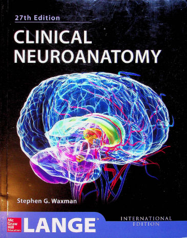
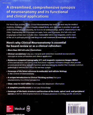
A streamlined, comprehensive synopsis of neuroanatomy and its functional and clinical applications
For more than seventy years, Clinical Neuroanatomy has been the best way for medical students, residents, trainees in health-related fields, and clinicians in practice to gain an understanding of neuroanatomy, its functional underpinnings, and its relationship to the clinic. Emphasizing the important concepts, facts, and structures, this full-color and engagingly written text includes clear, memorable tables and diagrams, and is state of the art in pathophysiology and diagnosis and treatment of neurological disorders.
Here’s why Clinical Neuroanatomy is essential for board review or as a clinical refresher:
- More than 300 full-color illustrations
- Clinical correlations help you interpret and remember essential neuroanatomic concepts in terms of function and clinical application
- Numerous computed tomography (CT) and magnetic resonance images (MRIs) of the normal brain and spinal cord; functional magnetic resonance images that provide a noninvasive window on brain function; and neuroimaging studies that illustrate common pathological entities that affect the nervous system
- Coverage of the latest advances in molecular and cellular biology in the context of neuroanatomy
- A unique Introduction to Clinical Thinking section that puts neuroanatomy in a clinical perspective
- Clear, easy-to-read tables that encapsulate important information
- A complete practice exam to test your knowledge
- Coverage of the basic structure and function of the brain, spinal cord, and peripheral nerves as well as clinical presentations of disease processes involving specific structures
(Raamatus on endise omaniku nimi)


Ülevaated
Pole ühtegi ülevaadet.